Home

Reorganiseren Voorganger brand cam 5.2 stain De volgende Zweet uitgehongerd

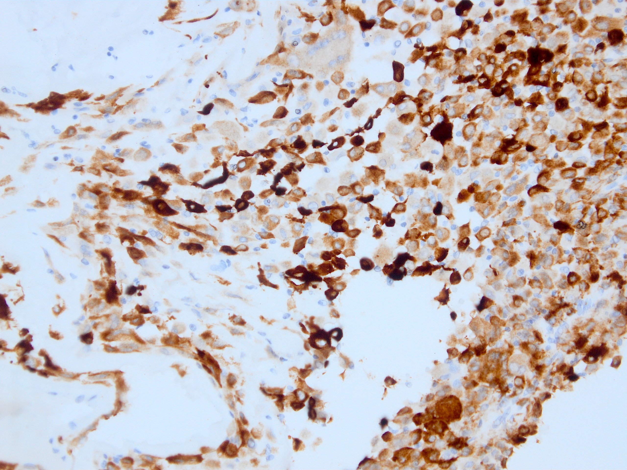
Cytokeratin (CAM5.2) - Mob469 - Diagnostic BioSystems(dbiosys) DBS

Positive cytokeratin cytoplasm staining CAM 5.2 in both populations... |  Download Scientific Diagram
Positive cytokeratin cytoplasm staining CAM 5.2 in both populations... | Download Scientific Diagram
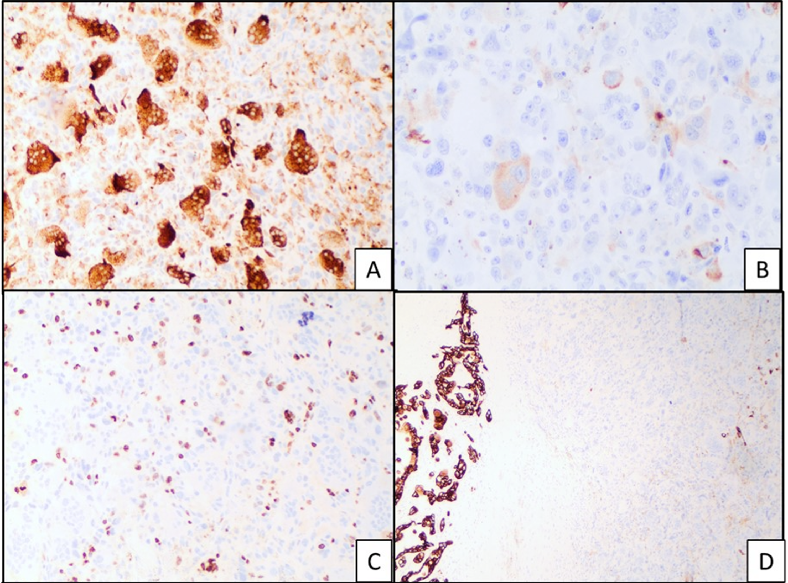
Macrophages and Dendritic Cells Are the Predominant Cells Infected in  Measles in Humans | mSphere
Macrophages and Dendritic Cells Are the Predominant Cells Infected in Measles in Humans | mSphere

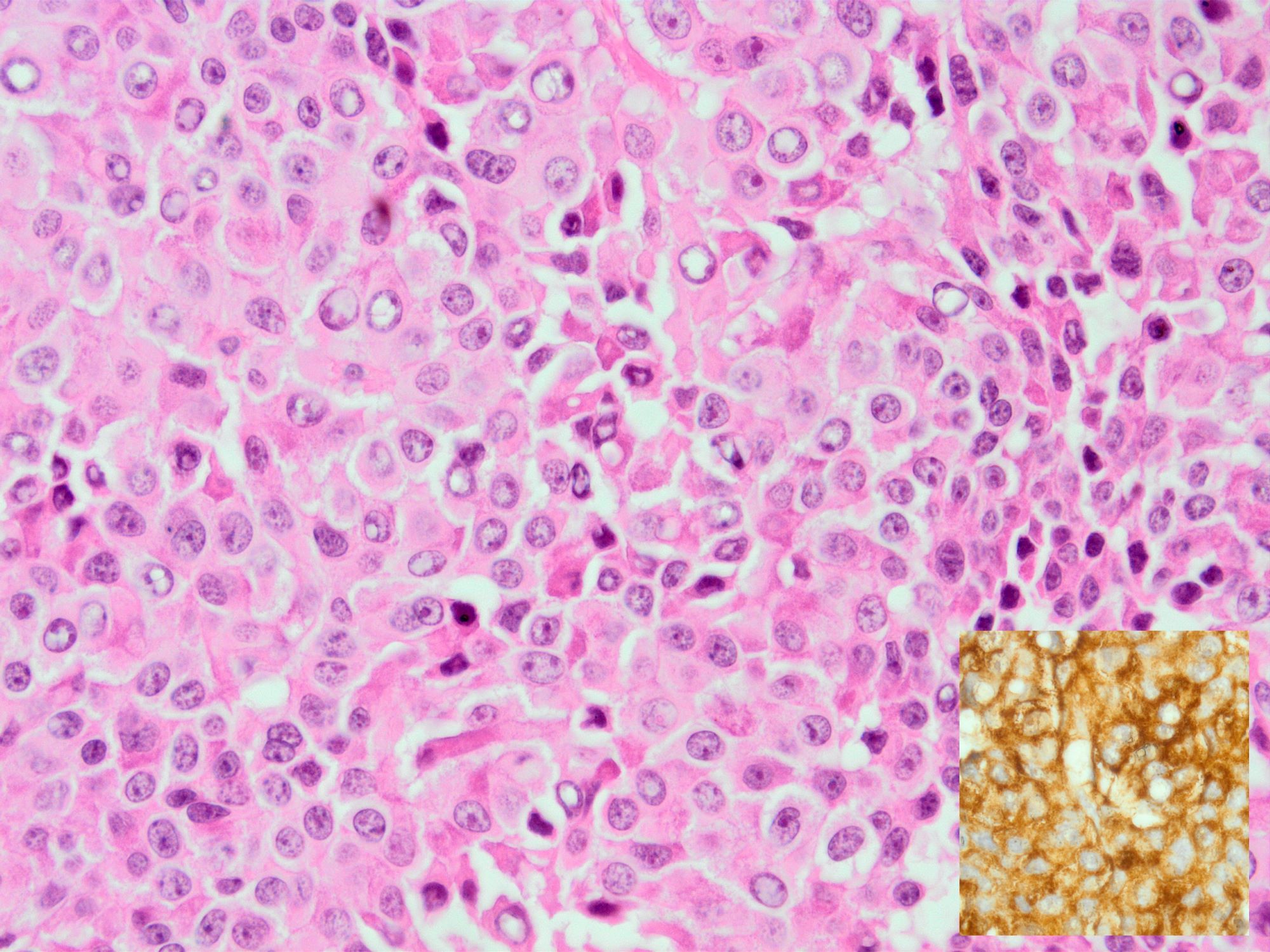
Pathology Outlines - Cytokeratin CAM 5.2

Immunohistochemistry in Normal Skin: Cytokeratins - YouTube

Skin Biopsy for Merkel Cell Carcinoma

Cureus | Undifferentiated Pancreatic Carcinoma With Osteoclast-Like Giant  Cells and Associated Ductal Adenocarcinoma With Focal Signet-Ring Features  | Article
Cureus | Undifferentiated Pancreatic Carcinoma With Osteoclast-Like Giant Cells and Associated Ductal Adenocarcinoma With Focal Signet-Ring Features | Article

Postmortem Investigation of Immunohistochemical Staining and Gross  Description of Sarcomatoid Carcinoma of the Lung in a Patient With Extreme  Leukemoid Reaction - Ali Ammar, Austin Ellis, Julia Hegert, Timothy W.  Jones, Rumi
Postmortem Investigation of Immunohistochemical Staining and Gross Description of Sarcomatoid Carcinoma of the Lung in a Patient With Extreme Leukemoid Reaction - Ali Ammar, Austin Ellis, Julia Hegert, Timothy W. Jones, Rumi
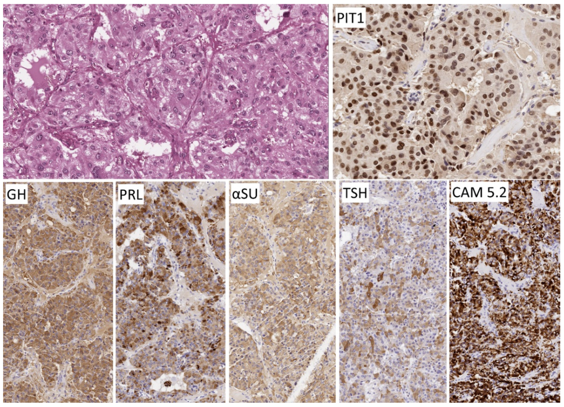
JCM | Free Full-Text | An Update on Pituitary Neuroendocrine Tumors Leading  to Acromegaly and Gigantism
JCM | Free Full-Text | An Update on Pituitary Neuroendocrine Tumors Leading to Acromegaly and Gigantism

Serial immunohistochemical stain for cytokeratin (CAM 5.2) and estrogen...  | Download Scientific Diagram
Serial immunohistochemical stain for cytokeratin (CAM 5.2) and estrogen... | Download Scientific Diagram

Thymoma - CAM 5.2 immunostain Case 243 | The epithelial cell… | Flickr

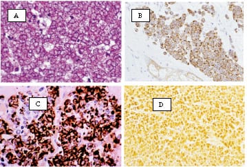
Cam5.2 staining of growth hormone adenoma. A. Densely granulated tumor... |  Download Scientific Diagram
Cam5.2 staining of growth hormone adenoma. A. Densely granulated tumor... | Download Scientific Diagram

Keratin-CAM5.2 | Pathnsitu Biotechnologies

Pathology Outlines - Cytokeratin CAM 5.2

Pigmented variation of Paget's disease with cytokeratin (CAM 5.2) staining  | Download Scientific Diagram
Pigmented variation of Paget's disease with cytokeratin (CAM 5.2) staining | Download Scientific Diagram

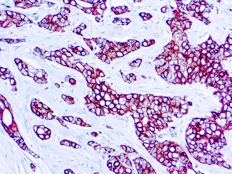
Innovative Science Press on Twitter: "Gastroblastoma is a rare biphasic  neoplasm of the gastric muscularis propria (usually antrum) that harbors a  MALAT1-GLI1 fusion. It sometimes behaves aggressively. The immunostain is a  keratin
Innovative Science Press on Twitter: "Gastroblastoma is a rare biphasic neoplasm of the gastric muscularis propria (usually antrum) that harbors a MALAT1-GLI1 fusion. It sometimes behaves aggressively. The immunostain is a keratin

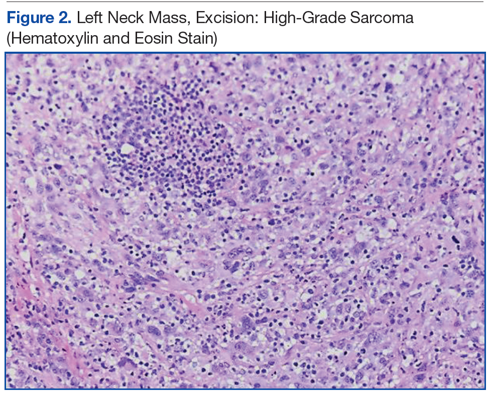
Complex Malignancies: A Diagnostic and Therapeutic Trilemma | AVAHO

Pathology Outlines - Cytokeratin CAM 5.2

Cytokeratin Family | Pathology Resident Wiki | Fandom

CAM 5.2 | 杭州百凌生物科技有限公司-官方网站

Cytokeratin LMW CAM5.2 Antibody (CAM5.2) - Bio SB

CAM 5.2 Stain Kit | Celltechgen LLC

Cytokeratin - CAM 5.2 | Pathology Resident Wiki | Fandom

Immunohistochemical Distinction of Ocular Sebaceous Carcinoma From Basal  Cell and Squamous Cell Carcinoma
Immunohistochemical Distinction of Ocular Sebaceous Carcinoma From Basal Cell and Squamous Cell Carcinoma